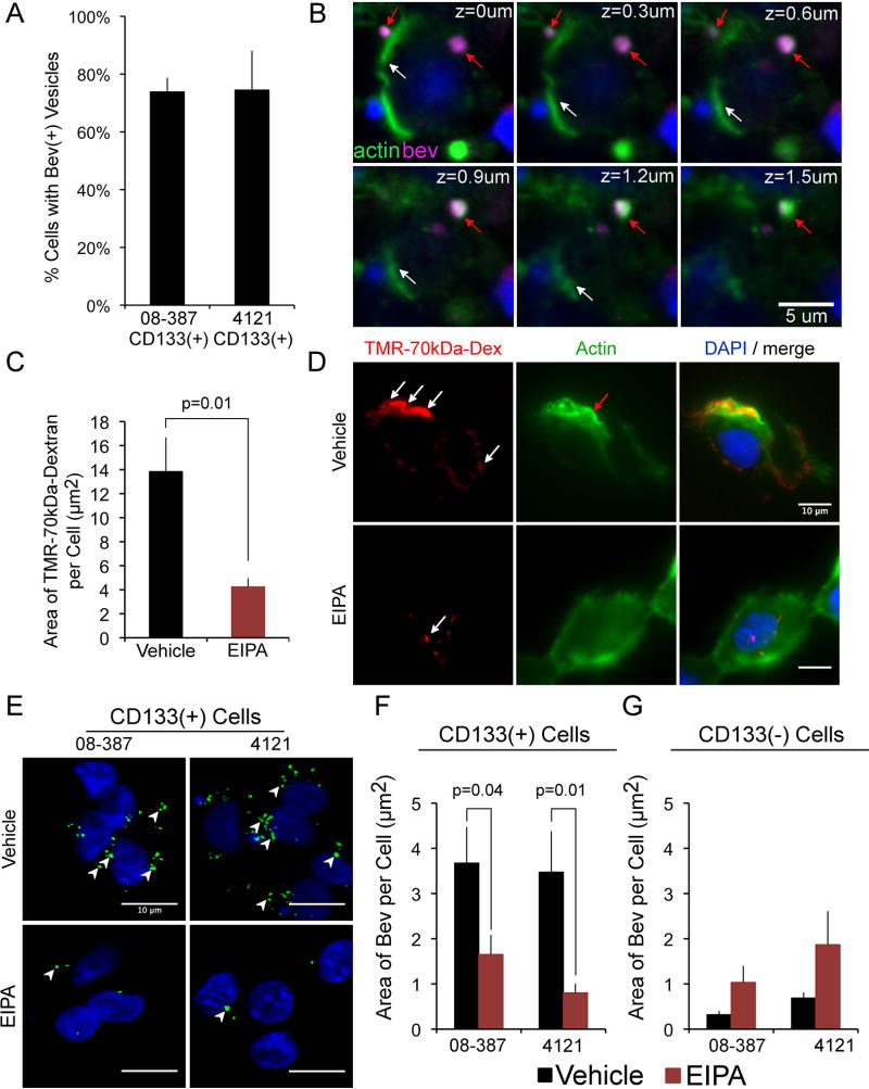
Figure 3.
Bevacizumab is internalized by CD133+ cells into membrane ruffles in a mechanism consistent with macropinocytosis. A&B, CD133+ cells were plated onto laminin in NBM without bFGF or EGF for 18 h (37°C, 5% CO2), followed by the addition of bevacizumab (250 µg/ml) for 5 min, the cells washed, fixed, reacted with Alexa-647-anti-human IgG (5ug/ml) and Alexa-488-phalloidin (5 units/ml), followed by DAPI nuclear stain, cover slipping, and confocal microscopy. Percent of CD133+ cells from two different tumor isolates (08–387 and 4121) containing bevacizumab-positive vesicles (A). A z-stack of the double-labeling is shown (CD133+ 08–387 cells) (B). White arrows denote phalloidin-stained cell membrane (green), and red arrows denote bevacizumab-positive vesicle (magenta) surrounded by actin. Scale bar denotes 5-µm. C&D, CD133+ cells (08–387) treated with 50 µM EIPA or vehicle for 30 min (37°C, 5% CO2), followed by addition of TMR-70-kDa-Dextran (1mg/ml) (red) for 5 min, were washed, reacted with Alexa-488-Phalloidin (green), nuclei stained with DAPI, cover slipped and microscopy performed. In cells treated with vehicle, TMR-70-kDa-Dextran is found in membrane ruffles (white arrows), containing polymerized actin (red arrows) (D). In cells treated with EIPA, reduced amounts of TMR-70-kDa-Dextran are internalized (C&D). The area (µm2) of TMR-70-kDa-Dextran/cell is plotted as the mean±SEM from >100 cells/condition (C). E–G, CD133+ GBM cells (08–387 and 4121) were plated on laminin in NBM as in panels A–D, and paired non-stem tumor cells (CD133-negative 08–387 and 4121) were plated in DMEM with 10% FBS for 18 h. CD133+ cells or the paired CD133-negative tumor cells were treated with vehicle or 50 µM EIPA for 30 min as above, followed by addition of bevacizumab (5 min), and the cells washed, fixed, reacted with Alexa-488-anti-human IgG, followed by DAPI nuclear stain, cover slipping and microscopy. Arrowheads denote bevacizumab-positive vesicles which are reduced in CD133+ cells treated with EIPA (E&F) but are not reduced in CD133-negative tumor cells treated with EIPA (G). The area (µm2) of bevacizumab/cell is plotted as the mean±SEM from >100 cells/condition (F&G). Scale bars denote 10-µm. Statistical analyses: exact two-sided Wilcoxon rank-sum tests.

