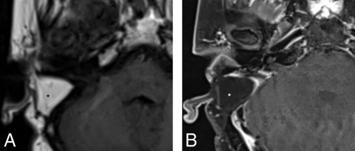
Fig 2.
Precontrast axial T1WI (A) and postcontrast axial T1WI with fat-suppression (B) demonstrate typical postoperative findings following a translabyrinthine craniotomy, with abdominal fat packing within the mastoidectomy defect (asterisk). Linear enhancement along the mastoidectomy bed reflects postsurgical changes without evidence of recurrent tumor within the IAC.

