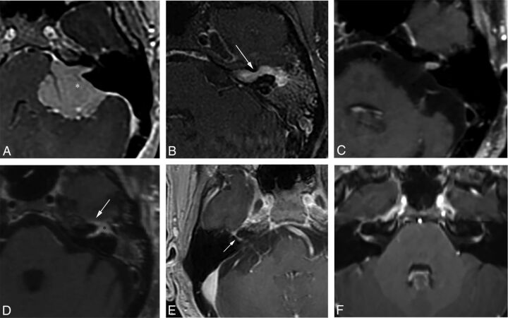
Fig 5.
Examples of various enhancing IAC and CPA masses on contrast-enhanced T1WI with fat-suppression (B–D, and F) and 3D echo-spoiled gradient-echo images (A and E). A, A large CPA meningioma, located eccentric to the porus acusticus (the asterisk denotes the tumor midline), extends into the IAC without the associated bony expansion often seen with VS (see Fig 6). B, An enhancing facial nerve schwannoma within the IAC extends into the labyrinthine segment (arrow), which differentiates a facial nerve from a vestibular schwannoma, as well as into the anterior genu and tympanic segments. C, A small enhancing metastatic lesion within the IAC in a patient with non-small cell lung cancer extends into the IAC fundus, labyrinthine, anterior genu, and tympanic segments. D, Perineural spread along the intratemporal and intracanicular segments of the facial nerve in a patient with squamous cell carcinoma of the periauricular skin (the asterisk indicates the anterior genu; arrow, the greater superficial petrosal nerve). E, An ill-defined tuft of enhancement within the IAC fundus extending into the labyrinthine segment (arrow) and anterior genu of the facial nerve in a patient with right Bell palsy. F, Bilateral ill-defined enhancement of the distal IAC bilaterally extending into the labyrinthine segment and anterior genu of the facial nerve canal in a patient with neurosarcoidosis.

