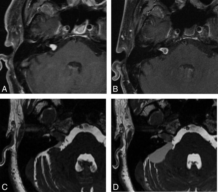
Fig 9.
Two examples of post-SRS imaging. Postcontrast axial T1WI with fat suppression in a patient before (A) and following (B) SRS reveals decreased enhancement centrally within the tumor on posttherapeutic imaging (B), confirming a positive response to SRS. Two axial FIESTA images (C and D) obtained during 2 consecutive follow-up examinations in a 2-year period demonstrate interval enlargement of the cystic component within the right CPA associated with a predominantly intrameatal VS following radiation therapy. The cystic component was later resected (not shown).

