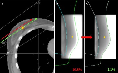
Figure 6.

Illustration of the effect of positioning errors on IVD results: (a) in presence of a setup error, the point of interest receives the correct dose but is associated with the wrong CT parameters, such as patient thickness; (b) the signal at the reference point is different than expected, thus a large dose difference is calculated; (c) by realigning the image with the TPS contours, the correct signal is reassigned to the reference point.
