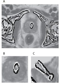
Figure 2.

The two contours are obtained from a bone and prostate stent segmentation in CT followed by an inverse transformation of the registration by the automatic (white) and the manual (black) registration for one patient. The segmentations are superimposed on the original MR images: (a) bones and prostate stent segmentation in the axial plane; (b) region of interest and the prostate stent in the axial plane; (c) region of interest and the prostate stent in the sagittal plane. It can be noticed, in particular in image (a), that both methods are based on a registration of the prostate stent and a tightly surrounding volume as either the black or white contour follow the contour of the pelvic bone. This results in an accurate alignment of the prostate stent in image (b), which indicates that the prostate has moved relatively to the pelvic bones. In image (c), a gap in the segmentation of the prostate stent can be observed from the automatic registration caused by interpolation.
