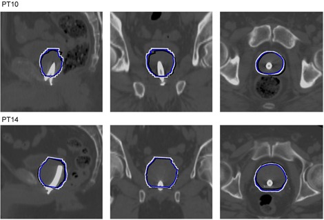
Figure 3.

The order is sagittal, coronal, and axial views of the overlap of the manual prostate delineation performed in the original MR image and then aligned to the CT based on the manual registration (black), the automatic registration of the CTV (blue), and the automatic registration of the extended CTV (white). The delineations are superimposed on the CT images.
