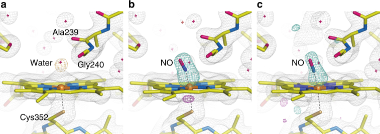
Fig. 4.
SFX structures of P450nor. a Resting-state structure. b, c Transient structures at 20 ms after caged-NO photolysis in the b absence and c presence of NADH. The 2F o−F c maps are shown in gray and contoured at 1.2σ. The F o−F c map is shown in orange and contoured at 4.0σ in a, whereas the F o(“Light”)−F o(“Dark2”) difference Fourier maps are shown in turquoise (positive) and magenta (negative) and contoured at 6.5σ in b and 3.2σ in c. All data were taken at ambient temperature. In a, the structure using the “Dark2” data of MC-2 is presented

