Fig. 2.
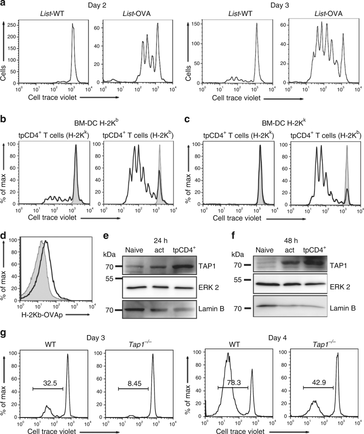
tpCD4+ T cells process bacterial antigens. a Proliferation of OT-I CD8+ T cells, 2 or 3 days after incubation with Listeria-WT or Listeria-OVA tpCD4+ T cells that captured bacteria from OVAp-I-loaded BM-DC. b Proliferation of OT-I CD8+ T cells incubated with Listeria-OVA tpCD4+ T cells (H-2Kk/k or H-2Kk/b), which captured bacteria from infected H-2Kb BM-DC, or with Listeria-WT tpCD4+ T cells (grey). c as for b, except transphagocytosis was carried out using infected H-2Kk/k BM-DC (48 h). d, H-2Kb/OVA expression (detected with anti-OVAp-I/H-2Kb antibody) by Listeria-OVA tpCD4+ T cells (H-2Kk/b, black line; H-2Kk/k, grey) that captured bacteria from H-2Kk/k BM-DC. e, f Western blot showing TAP1 expression in naive, activated (act, by OVAp-I-loaded BM-DC) CD4+ T cells, or Listeria-OVA tpCD4+ T cells at 24 e or 48 h f after activation. ERK2 and laminB were used as protein loading controls. Note that all original western blots together with the membranes stained for molecular size markers are provided at the end of the manuscript in Supplementary Fig. 5. g Proliferation of CD8+ T cells from OT-I mice, 3 or 4 days after conjugation with Listeria-OVA tpCD4+ T cells (from Tap1−/− or isogenic WT mice)

