Figure 2.

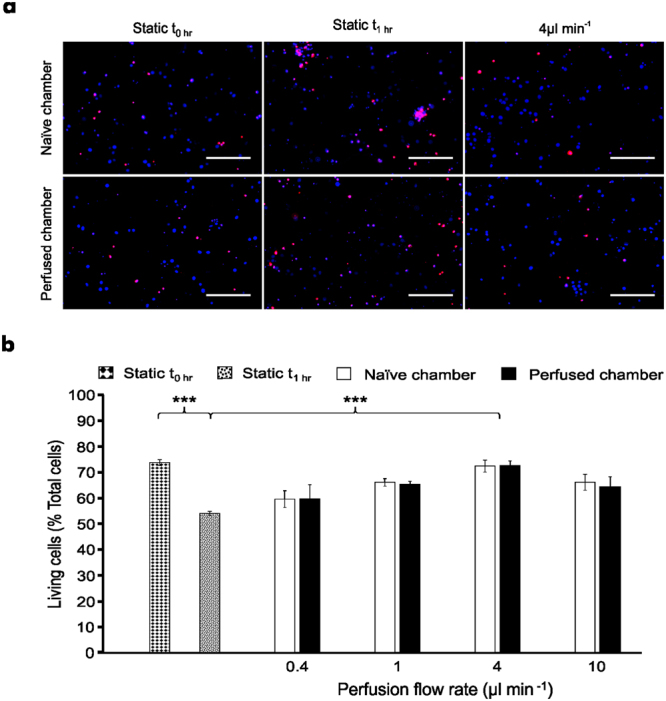
Continuous perfusion maintains cell viability between environmentally isolated neural networks. (a) Representative images obtained from perfused and naïve cultures which were stained with Hoechst (blue) and PI (red) following continuous perfusion experiments for 1 hour. Scale bar = 200 µm. (b) Microfluidic neural network viability increases in the presence of perfusion with respect to static condition after 1 hour, with an optimal flow rate of 4 μl min−1. Data are presented as mean ± S.E.M. with one-way ANOVA with post-hoc Tukey’s test performed; n = 21 devices from 4 cultures; ***denotes P < 0.001.
