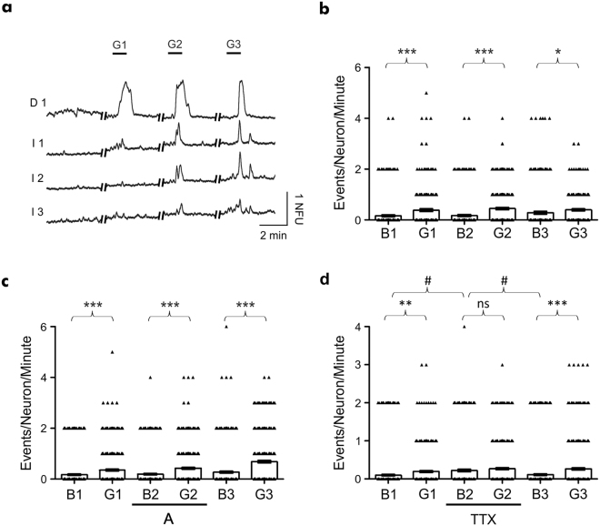
Figure 5.
Repeated glutamate application induces increased neuronal activity in synaptically connected hippocampal cultures. (a) Ca2+ imaging traces representative of activity obtained from individual neurons in the directly perfused (trace D1) and naïve (traces I1–I3) culture chambers, demonstrating functional synaptic communication between independent hippocampal cultures. (b) Significant increases in the number of neuronal calcium events were observed in the naïve chamber, with respect to the basal (B) activity, in response to the three glutamate (G) applications in the perfused chamber. (c) Significant increases in the number of neuronal calcium events were observed in the naïve chamber, with respect to the basal (B) activity, in response to glutamate applications (G) in the absence and presence of glutamate antagonists in the perfused chamber. (d) Basal activity in the naïve chamber was significantly increased in the presence of TTX in the perfused chamber. Further changes in the number of neuronal calcium events were not observed following glutamate (G) application in the presence of TTX. Data are presented as mean ± S.E.M. with paired student’s t-test performed; n = ≥ 222 neurons per application, ≥3 devices from ≥3 separate cultures; ns denotes P > 0.05, *denotes P < 0.05, **denotes P < 0.01, ***denotes P < 0.001 and #denotes P < 0.05.

