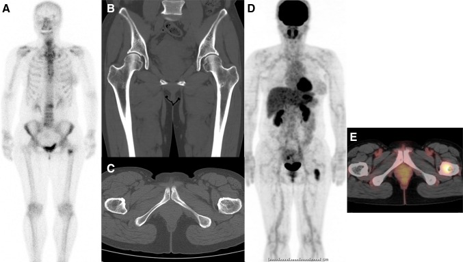
Fig. 1.
A 51-year-old female had right breast cancer surgery 10 years ago. She showed a tumor marker elevation (carcinoembryonic antigen), and bone scintigraphy (BS) was performed. A hot spot was shown in her left femur (around lesser trochanter) on BS (a). However, following CT could not demonstrate the lesion (b coronal, c axial). Therefore, FDG-PET/CT study was carried out, and an intense FDG uptake was noticed on her left femur (d whole-body FDG image, e fusion axial image). CT-guided bone biopsy revealed the lesion as metastasis pathologically

