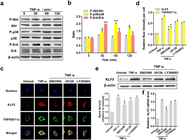
Figure 6.
TNF-α induces KLF5 expression via the p38 pathway in cervical cancer cells. (a) Erk, Akt and p38 MAPK phosphorylation in HeLa cells was analysed by western blotting. β-actin was used as the loading control. (b) The activation levels of these three kinases were determined by the respective phosphorylation ratios. *P < 0.05, **P < 0.01. (c) The cellular locations of KLF5 (red) and tumour necrosis factor receptor superfamily member 11a (TNFRSF11a, green) in HeLa cells were determined by immunofluorescence staining (×200). DAPI was used for nuclear staining. Scale bars = 10 μm. (d) Statistical analysis of fluorescence intensity. *P < 0.05 vs. the TNF-α group. KLF5 expression in HeLa cells was determined at the protein (e) and mRNA levels (f). *P < 0.05 vs. the TNF-α group.

