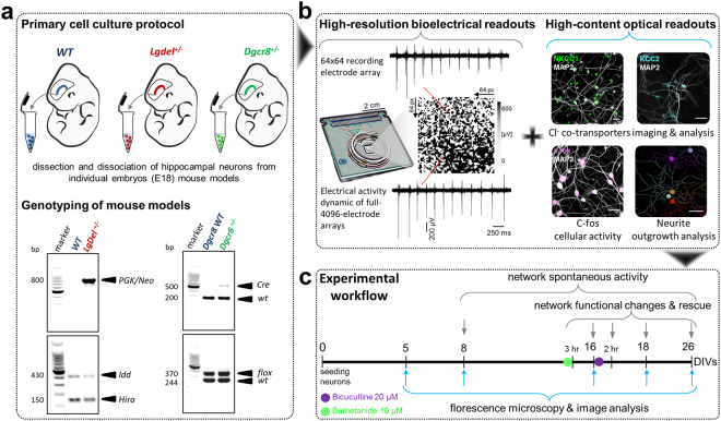
Figure 1.
Schematic of experimental implementation of the 22q11.2 DS model. (a) Single-embryo primary cell culture preparation of hippocampal neurons from WT, Lgdel +/− and Dgcr8 +/− mice and their genotyping. Each cropped gel image reports two representative embryos from groups of animals genotyped for PGK/Neo (top left) and Idd/Hira alleles (bottom left) in (WT and Lgdel +/− embryos) or Cre (top right) and flox alleles (bottom right) in (Dgcr8 WT and Dgcr8 +/− embryos); “full-length gels are presented in Supplementary Figure S5”. (b) Overview of the experimental readouts used in this study, combining electrical measures and optical HCI. Scale bars represent 30 μm. (c) Workflow timeline of the experimental implementation.

