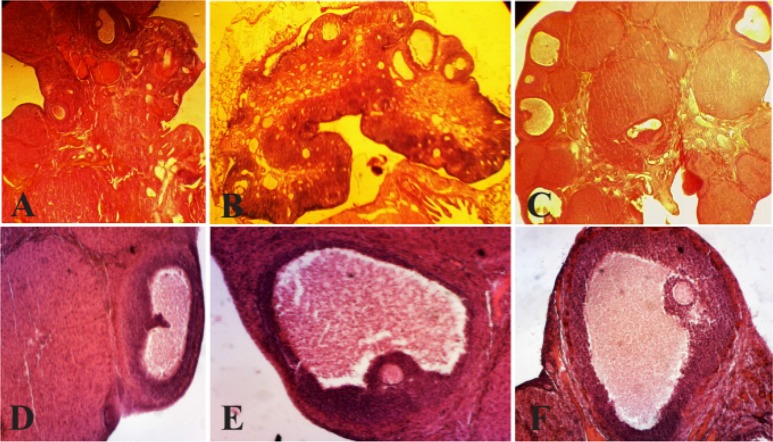
Figure 1.
Histological analysis of normal ovaries (A, D) compared with PCOS (B, E) and ovaries treated with curcumin (C, F). The morphological changes of the rats’ ovarian tissues were stained with hematoxylin and eosin, as described in the Materials and Methods section. A, D) A representative rat’s ovarian tissue section from the control group, which had normal appearance (A×100,40). B, E) A representative rat’s ovarian tissue section from the PCOS group showed thickening surface albuginea, under which there were many follicles in different phases (including atretic follicles and cystic dilating follicles), as well as fewer layers of granular cells, disappeared oocytes and corona radiating within the follicles (B×100, 40). C, F) A representative rat’s ovarian tissue section from the group treated with curcumin, which showed increased granular cell layers, and some ovulation phenomena (C×100, 40) (Scale bar, 50 μm A, B, C), (Scale bar, 20 μm D, E, F). AF: atretic follicle, CF: cystic follicle (*), CL: corpus luteum, GCL: granular cell layer (Δ), TCL: theca cell layer (→)

