Figure 4.
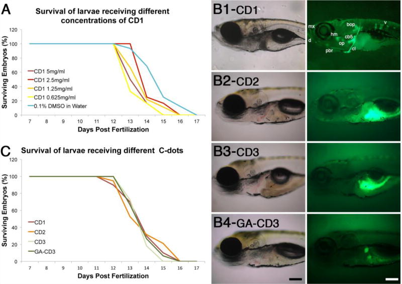
Carbon dots synthesized from carbon powder (CD1) are non-toxic and unique in their ability to bind bone. (A) Survival curves of larvae injected with different amounts of CD1. (B) Transmitted and fluorescent images of 8-day old larvae injected at 6 days post fertilization with C-dots synthesized from carbon powder (B1, CD1), citric acid (B2, CD2), glycerin (B3, CD3), and glutamic acid-conjugated glycerin derived C-dots (B4, GA-CD3). In B1, bones are dentary (d), maxilla (mx), posterior branchiostegal ray (pbr), hyomandibula (hm), opercle (op), ceratobranchial 5 (cb5), cleithrum (cl), basioccipital articulatory process (bop), and vertebrae (v). Images in B2 and B3 were over-exposed to demonstrate that CD2 and CD3 do not bind to bones; the autofluorescence seen in the gut tissues is a result of image over exposure. In B4, the two stained structures correspond to primitive kidneys (pronephros). Scale bar is 100 microns. (C) Survival curves of larvae injected with different C-dots preparations. For both graphs, survival curves for control (0.1% DMSO in water) and C-dots injected larvae are not significantly different (n≥25 embryos per condition in at least two independent experiments).

