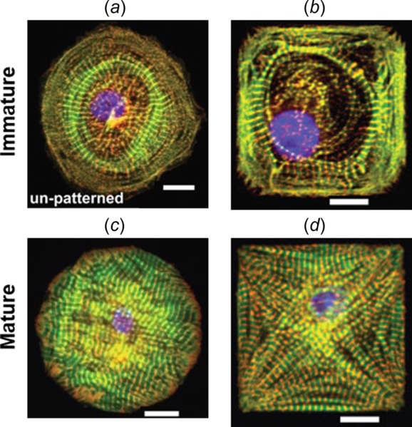
Fig. 2.

Different myofibril organizations observed in (a) and (b) immature (stem-cell derived) versus (c) and (d) mature (neonatal) cardiomyocytes for the same cell shapes. (Reprinted with permission from Sheehy et al. [6]. Copyright 2012 by Springer.)
