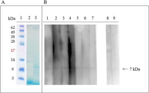
Figure 2.

Patient IgE binding to Japanese apricot gibberellin‐regulated protein detected by IgE‐Immunoblotting. (A) Coomassie blue staining of PVDF membrane: lane 1, molecular mass standard; lane 2, Japanese apricot protein extract; lane 3, purified Japanese apricot gibberellin‐regulated protein. (B) IgE‐immunoblotting using purified Japanese apricot gibberellin regulated protein: lanes 1–7, patients 1–7; lanes 8–9, control individuals.
