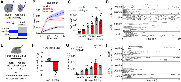
Figure 8. AgRP neurons are epistatic to leptin's effect on food intake.
(A) Schematic of experiments in (B-D). Optogenetic stimulation of AgRP neurons in ad libitum fed WT and ob/ob mice prior to (prestim) or during (costim) food availability. Blue indicates the timing of laser stimulation.
(B) Cumulative food intake by ob/ob mice after no stimulation (black), 60 min pre-stimulation (red), or during 60 min co-stimulation (blue). Traces represent mean ± SEM (n = 6-10 mice per group).
(C) Quantification of food intake from (B).
(D) Raster plots showing feeding pattern in individual mice from (B and C).
(E) Schematic of experiments in (F-H). Optogenetic stimulation of AgRP neurons in ad libitum fed, ob/ob mice during chronic vehicle or leptin infusion by mini-osmotic pumps. Stimulation occurred prior to (prestim) or during (costim) food availability as in (A).
(F) Bodyweight change in ob/ob mice 3 days after implantation of a mini-osmotic pump infusing vehicle (gray, n = 6 mice) or leptin (red, n = 7 mice)
(G) Quantification of food intake by ad libitum fed ob/ob mice from (F) receiving chronic vehicle (gray) or leptin (red) infusion after no stimulation, 60 min pre-stimulation, or during 60 min co-stimulation.
(H) Raster plots showing feeding pattern in individual mice from (G).
(C, F, G) ■ denotes individual mice. Bars represent mean ± SEM. *P < 0.05, **P < 0.01, ***P < 10-3. (D and H) Each row represents a single trial from a different animal and each point indicates consumption of a 0.02 g pellet.
See also Figure S7.

