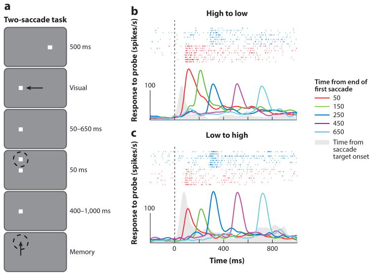
Figure 11.
Postsaccadic inaccuracy of gain fields in the lateral intraparietal area. (a) The two-saccade task. Dashed circle represents the receptive field of the neuron under study, and arrows represent directions of saccades. Single-cell responses to probes flashed at different times after a conditioning saccade in the (b) high-to-low and (c) low-to-high directions. Activity immediately following the conditioning saccade consistently indicates the presaccadic eye position. Activity is aligned on the end of the first saccade (dotted line), averaged across trials, and convolved with a 20-ms Gaussian filter. Colors indicate different timings of the probe. Rasters at top in panels b and c show spikes in the 50-ms (red) and 250-ms (blue) probe delay conditions. The solid curve (gray) shows the steady-state visual response at the postsaccadic orbital position during a memory-guided saccade task; for this curve, zero on the abscissa is the time of appearance of the saccade target. Figure adapted from Xu et al. (2012) with permission.

