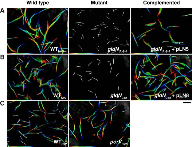
FIG 4.
Gliding of wild-type and mutant cells on glass. Cells were grown in TYES (A) or Shieh medium (B and C) at 28°C for 14 h (early stationary phase). Ten-microliter aliquots of cultures were introduced into tunnel slides and observed for motility using an Olympus BH-2 phase-contrast microscope with a heated stage set at 25°C. (A) WT F. columnare IA-S-4, ΔgldN mutant of strain IA-S-4 (ΔgldNIA-S-4), and the ΔgldNIA-S-4 mutant complemented with wild-type gldN on pLN5. (B) Wild-type F. columnare C#2, ΔgldN mutant of strain C#2 (ΔgldNC#2), and the ΔgldNC#2 mutant complemented with wild-type gldN on pLN8. (C) Wild-type F. columnare C#2 and the ΔporV mutant of strain C#2 (ΔporVC#2). In each case, a series of images were taken for 20 s using a Photometrics CoolSNAP cf2 camera. Individual frames were colored from red (time zero) to yellow, green, cyan, and finally blue (20 s) and integrated into one image, resulting in “rainbow traces” of gliding cells. The rainbow traces correspond to the first 20 s of the sequences shown in Movie S1 (top row [A]), Movie S2 (middle row [B]), and Movie S3 (bottom row [C]). White cells correspond to cells that exhibited little if any net movement. The first frame of each movie (time zero) is shown in Fig. S1. Scale bar at lower right indicates 10 μm and applies to all panels.

