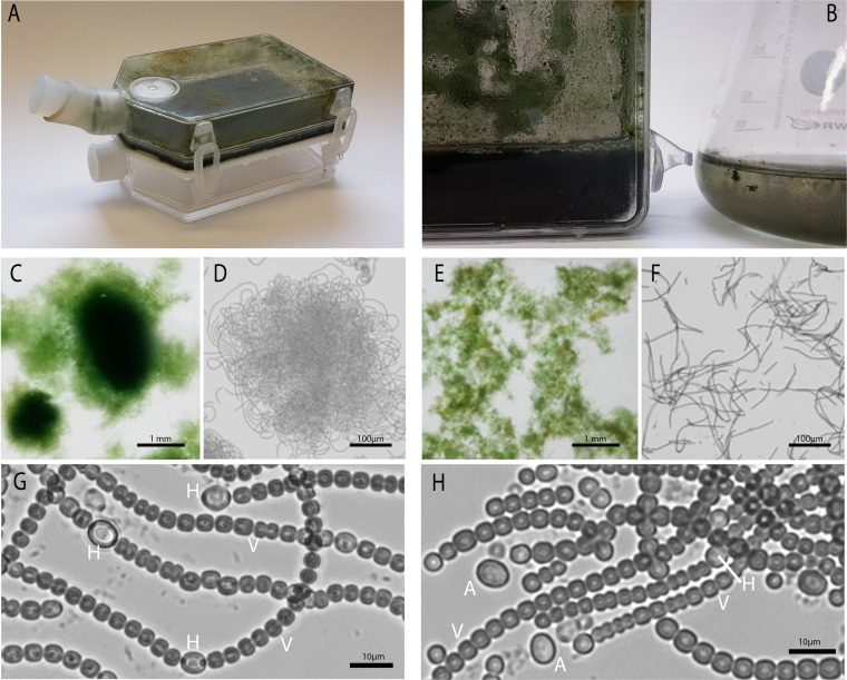
FIG 2.
Macroscopic and microscopic appearances of N. punctiforme PCC 73102 in HD and C batch cultures. (A) Two-tier growth vessel with thick biofilm of N. punctiforme. (B) Comparison of cell densities in HD (left) and C (right) cultivation. (C) Tufts of N. punctiforme observed in HD cultures. (D) Microscopic overview of N. punctiforme filament aggregation in HD cultures. (E) Cluster of filaments of N. punctiforme observed in C cultures. (F) Loose packing of filaments of N. punctiforme in C cultures. (G and H) Cell types observed in HD and C cultures, respectively. Labels: V, vegetative cell; H, heterocyst; A, enlarged single cell.

