Figure 4.

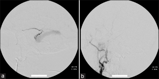
(a) Selective micro catheterization of the left middle meningeal artery showing the fistulous point. (b) Final control of the second session, after embolization of the left middle meningeal artery

(a) Selective micro catheterization of the left middle meningeal artery showing the fistulous point. (b) Final control of the second session, after embolization of the left middle meningeal artery