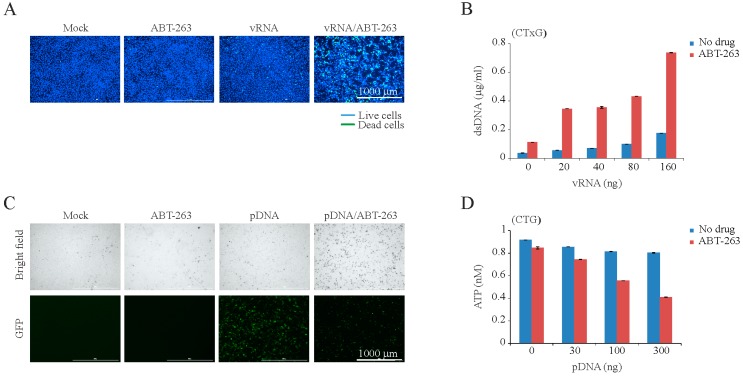
Figure 3.
ABT-263 induces the premature death of cells transfected with IAV genomic RNA (vRNA) or plasmid DNA (pDNA). (A) Fluorescent microscopy images showing that ABT-263 kills vRNA-transfected (160 ng) but not mock-transfected RPE cells at 8 h post transfection. Asymmetric cyanine dye stains the dsDNA of dead cells. Hoechst stains DNA in living cells; (B) CTxG plot showing that ABT-263 (3 µM) induces that premature death of RPE cells transfected with increasing concentrations of vRNA. Mean ± SD, n = 3; (C) Fluorescent and bright field microscopy of RPE cells showing that ABT-263 kills eGFP-expressing plasmid transfected (300 ng) but not mock-transfected RPE cells at 6 h post transfection; (D) CTG graph showing that the viability of ABT-263-treated (3 µM) cells decreases with increasing concentrations of transfected plasmid DNA. Mean ± SD, n = 3.

