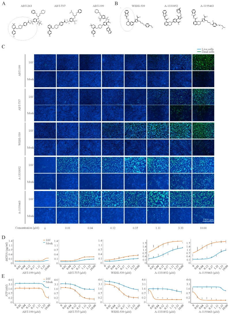
Figure 4.
Anti-IAV activities of ABT-263 analogues. (A,B) Structures of ABT-263, ABT-737, and ABT-199, as well as WEHI-539, A-1331852, and A-1155463, showing that these molecules share similar elements; (C) fluorescent microscopy images showing that increasing concentrations of Bcl-2i kill IAV-infected (moi 3) but not mock-infected RPE cells at 24 hpi. Asymmetric cyanine dye stains the dsDNA of dead cells. Hoechst stains DNA in living cells; (D) quantification of dsDNA in dead cells. Mean ± SD, n = 3; (E) quantification of intracellular ATP in living cells using CTG assay. Mean ± SD, n = 3.

