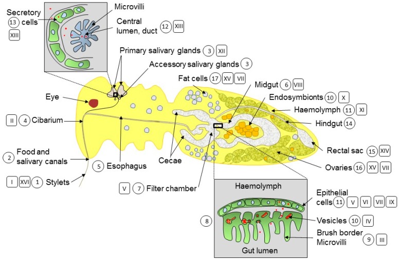
Figure 1.
Schematic drawing of a female whitefly longitudinal cross-section. The main organs and cells involved in begomovirus translocation (Arabic numerals in a circle) and the main processes affecting the viruses (Roman numerals in a rectangle) are shown in the drawing. The important virus translocation sites in the midgut and the primary salivary glands are enlarged. Organ and cells: 1. stylets; 2. maxillary stylet containing the food and salivary canals; 3. primary and accessory salivary glands; 4. cibarium; 5. esophagus; 6. filter chamber; 7. midgut; 8. section across the filter chamber; 9. microvilli; 10. bacteriosome and endosymbiotic bacteria; 11. haemolymph; 12. salivary gland lumen; 13. salivary gland secretory cells; 14. hindgut; 15. rectal sac; 16. ovaries; 17. fat cells. Processes: I. virus ingestion; II. cibarium: discrimination non-circulative and circulative viruses; III. entry of virus via microvilli; IV. endocytosis; V. virus accumulation; VI. transcription, replication, virion formation; VII. autophagy; VIII. interactions virus CP-whitefly proteins; IX. virus aggregation; X. GroEL production; XI. interaction endosymbiotic GroEL-begomovirus CP; XII. release of virion from GroEL near primary salivary glands; XIII. secretion of begomovirus in primary salivary glands, from secretory cells into central lumen; XIV. virus excretion with honeydew; XV. virus invasion of fat cells and ovaries; XVI. virus egestion and transmission to plants.

