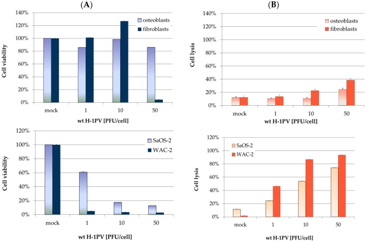
Figure 2.
Innocuousness of wild-type H-1PV towards non-transformed mesenchymal cells. (A) 3-(4,5-dimethylthiazol-2-yl)-2,5-diphenyltetrazolium bromide (MTT) test for cell viability quantification, performed 7 days after H-1PV infection. Metabolic activity values were normalized to mock-treated control cells. Bars represent means of eight independent experiments, and the corresponding error bars represent standard errors of the mean (SEM); (B) lactate dehydrogenase (LDH)-release assays for quantifying H-1PV-induced cell lysis. LDH release was assayed three days after H-1PV infection. LDH activity in supernatants was normalized with respect to complete lysis with Triton. SaOS-2 and WAC-2 (lower panel) were used as positive controls.

