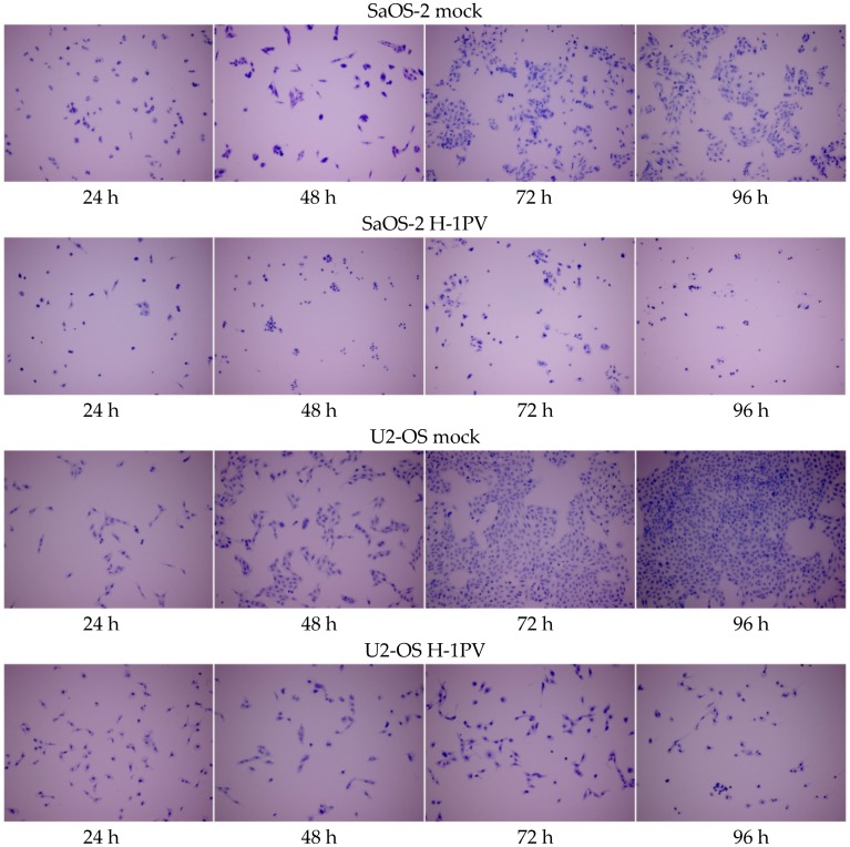
Figure 4.
H-1PV infection induces antiproliferative and cytotoxic effects in osteosarcoma cell lines. Microscope images of osteosarcoma cells stained with crystal violet at 24-h intervals for up to 96 h after infection with 1 PFU wild-type H-1PV per cell. Upper panels: mock-infected cells. Magnification: 100×.

