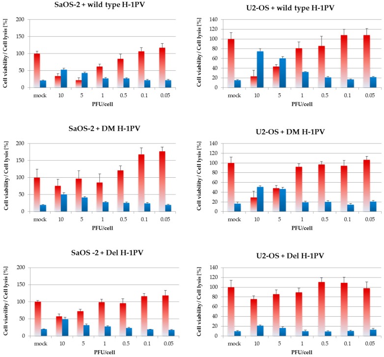
Figure 7.
Six days after infection, wild-type H-1PV shows stronger toxicity towards osteosarcoma cells than mutant viruses. Cells were infected with increasing titers of wild-type H-1PV, Del H-1PV, or double-mutant H1-DM. Six days after infection, cytotoxicity testing was performed in 96-well plates in simultaneous MTT tests and LDH-release assays. Red and blue bars represent means of eight independent experiments and error bars represent standard errors of the mean (SEM). Cell viability is indicated in blue and cell lysis in red.

