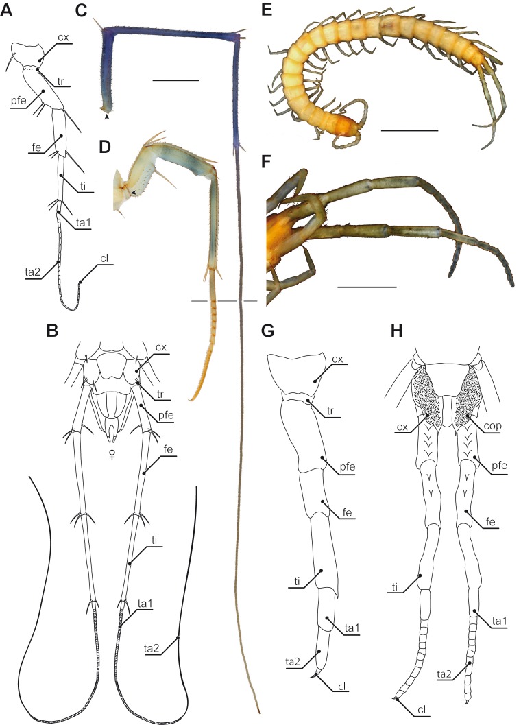
Figure 3. Ultimate legs in Scutigeromorpha and Newportia spp.
(A) Schematic representation of walking leg 10 of Scutigera coleoptrata. The breaking point (for autotomization) of the leg is located between trochanter and prefemur, indicated by a dotted line (compare black arrowhead in C and D, Original). (B) Schematic representation of the posterior trunk and ultimate legs of a female S. coleoptrata (view from ventral, modified after Minelli & Koch, 2011). (C) Ultimate leg of S. coleoptrata (compare B, Original). Tip of tarsus 2 is incompletely regenerated. Transition of tarsus 1 and 2 indicated by grey line. (D) Walking leg 10 of S. coleoptrata equally scaled to the ultimate leg (compare A, Original). Transition of tarsus 1 and 2 indicated by grey line. (E) Habitus of Newportia monticola Pocock, 1890 (view from dorsal, Original). Note the comparable length of antennae and ultimate legs. (F) Close up of ultimate legs with multi-annulated tarsus of N. monticola (view from ventral, compare H, Original). (G) Schematic representation of walking leg 10 of Newportia longitarsis (Newport, 1845) and (H) Schematic representation of the posterior trunk and ultimate legs of N. longitarsis (view from ventral, compiled after Schileyko & Minelli, 1998). Scale bars: A, C 500 µm, E 250 µm, F 100 µm. Abbreviations: cl, pretarsal claw; cx, coxa; fe, femur; pfe, prefemur; ta1, tarsus 1; ta2, tarsus 2; ti, tibia; tr, trochanter.

