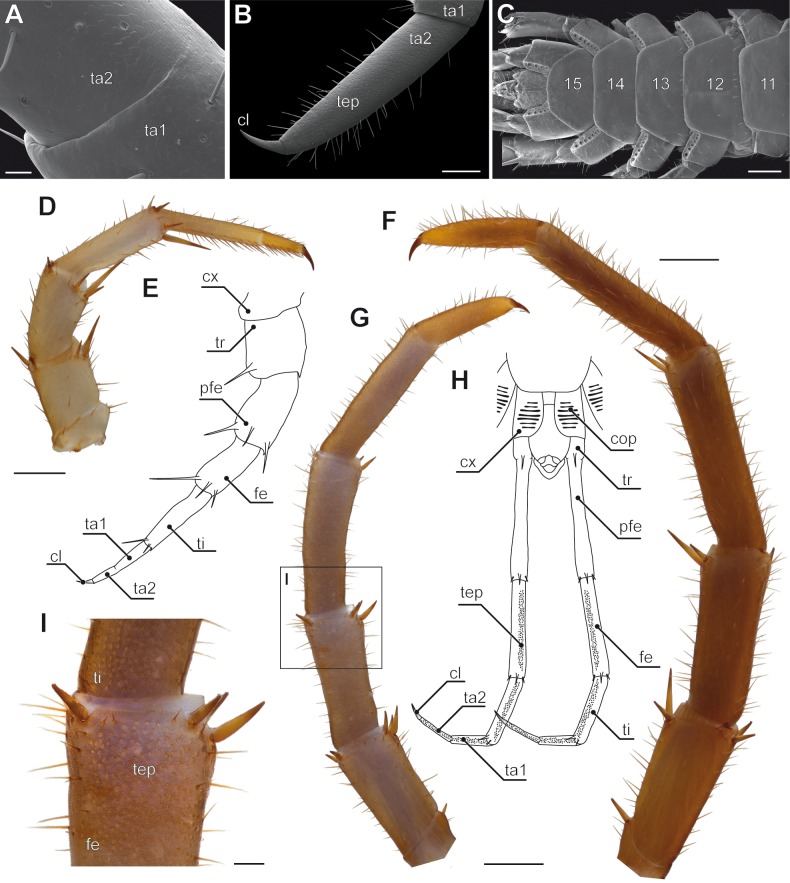
Figure 4. Aspects of ultimate legs in Lithobius forficatus.
(A) Incomplete separation of tarsus 1 and 2 of the walking leg 10 (SEM, Original). (B) Tarsus 2 and pretarsal claw of the ultimate leg, medial side with pores of telopodite glands (SEM, Original). (C) Posterior trunk from ventral. Coxae of legs 12 to 15 (ultimate leg) each possess a row of coxal pores (SEM, Original). (D) Walking leg 10. Note the incomplete separation of tarsus 1 and 2 indicated by the whitish interpodomeric cuticular membrane (Original). (E) Schematic representation of walking leg 10 (Original). (F) Left ultimate leg from lateral (Original). (G) Right ultimate leg from medial (Original). Note the milky appearance of the cuticular surface due to the pores of telopodite glands. (H) Schematic representation of the posterior trunk with ultimate legs (view from ventral, modified after Minelli & Koch, 2011). (I) Inset as indicated in G showing the pores of telopodite glands on femur and tibia. Scale bars: A 20 µm, B 200 µm, C, D, G, F 500 µm, I 100 µm. Abbreviations: cl, pretarsal claw; cop, coxal pores; cx, coxa; fe, femur; pfe, prefemur; ta1, tarsus 1; ta2, tarsus 2; tep, pore of telopodite glands; ti, tibia; tr, trochanter, 11–15 sternites of leg-bearing segments 11 to 15.

