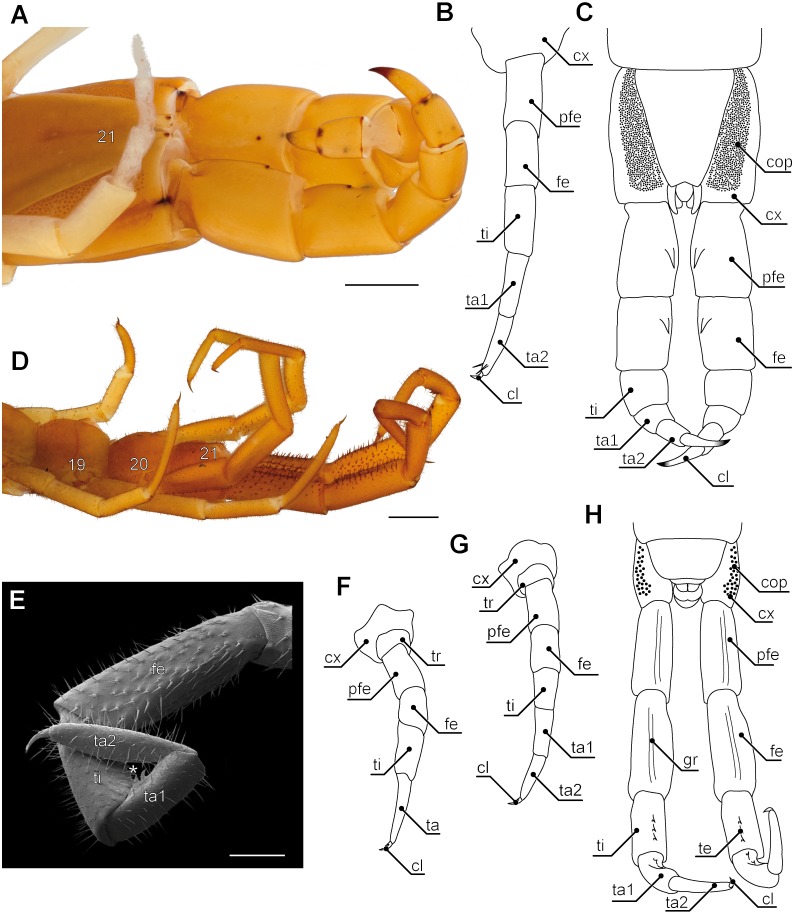
Figure 5. Aspects of ultimate legs in Theatops spp. and Cryptops hortensis.
(A) Ultimate legs of Theatops erythrocephalus C.L. Koch, 1847 (view from ventrolateral, Original). Tarsal podomeres are slightly detached. The ventral coxopleura is covered with coxal pores. (B) Schematic representation of walking leg 10 in T. erythrocephalus (Original), and (C) the posterior trunk with ultimate legs in Theatops californiensis Chamberlin, 1902 (modified after Shelley, 1997). (D) Posterior trunk and walking legs 19, 20, as well as ultimate legs of Cryptops hortensis (view from ventrolateral, Original). (E) Left ultimate leg “claw” of C. hortensis (view from medial, SEM, Original). Note the tibial and tarsal ‘sawteeth’ (asterisk). Please note the conflicting terminology of podomeres and compare also Fig. 6A. (F–G) Schematic representation of walking leg 19 (F) and walking leg 20 (G). Note the division of the tarsus in walking leg 20 (Originals). (H) Schematic representation of the posterior trunk and ultimate legs (view from ventral, Original). Note the ventral grooves on prefemora and femora, as well as the ‘teeth’ on tibia and tarsus 1. Scale bars: A 1 mm, D 500 µm, E 250 µm.Abbreviations: cl, pretarsal claw; cop, coxal pores; cx, coxa; fe, femur; gr, grooves on prefemora and femora; pfe, prefemur; ta, tarsus; ta1, tarsus 1; ta2, tarsus 2; te, ‘teeth’ of tibia and tarsus 1; ti, tibia; tr, trochanter; 19–21 sternites of leg-bearing segments 19 to 21.

