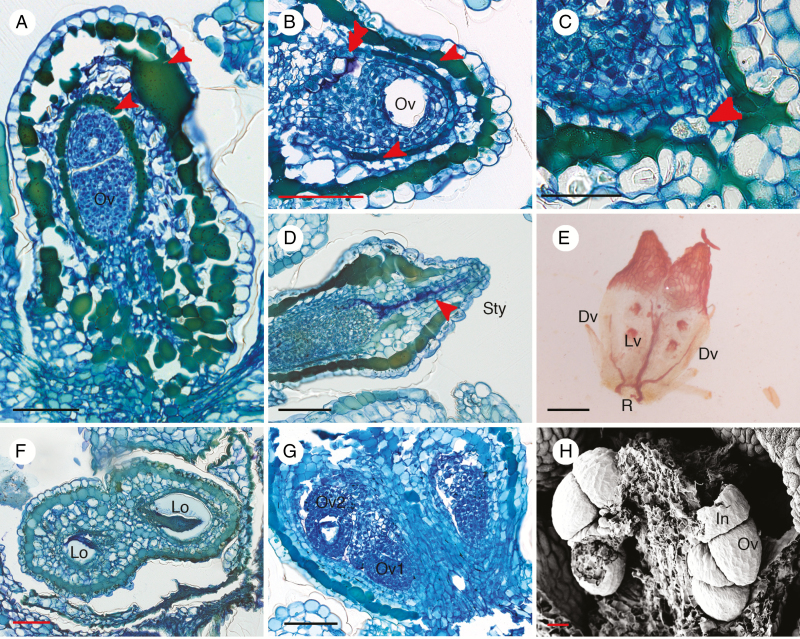
Fig. 5.
Anatomical structures of Ophiocaryon gynoecium. (A–D, G, H) O. heterophyllum; (E) O. paradoxum; (F) O. duckei. (A) Longitudinal section of ovary presents two layers of tanniferous cells, in the hypodermis and endodermis (arrowheads); two superposed ovules are visible inside a locule. (B) Transverse section of ovary showing an ovule inside a locule; there are two layers of tanniferous tissue in the ovary wall (arrowheads); note purple intralocular hairs (double arrowhead). (C) Presence of calcium oxalate crystal at the base of the ovary (arrowhead). (D) Longitudinal section of young ovary with secretion visible in the stylar canal (arrowhead). (E) Cleared ovary showing vascular bundles forming a ring (R) at the base and branching into two Y-shaped lateral veins (Lv) and two dorsal veins (Dv); no vascular bundles reach into the style. (F) Transverse section of an ovary consisting of two carpels with axile placentation; ovules are alternately attached on different sides of the locule. (G) Two superposed ovules are present in each locule. (H) Scanning electron microscopic image of young ovules demonstrating that integuments of ovules are reduced and swollen. Dv, dorsal vein; In, integument; Lv, lateral vein; Lo, locule; Ov, ovule; R, vascular ring; Sty, style. Scale bars: (A–D, F, G) = 100 μm; (E) = 200 μm; (H) = 30 μm.

