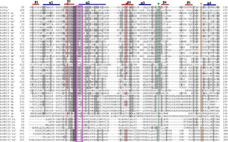
Fig. 3.

Multiple sequence alignment of the a-type domains of B. rapa PDI proteins and a typical human PDI. Residues highlighted in gray and light gray share 100% and >50% identity, respectively. Elements of the secondary structure are specified by blue colour bars (α-helices) and red colour bars (β-sheets). Red arrows indicate the two buried charged residues in the vicinity of the active site, orange arrow indicates the conserved arginine (R) and green arrow indicates the cis prolines (P) near each active site. Active-site residues within a domain are pink colour boxed
