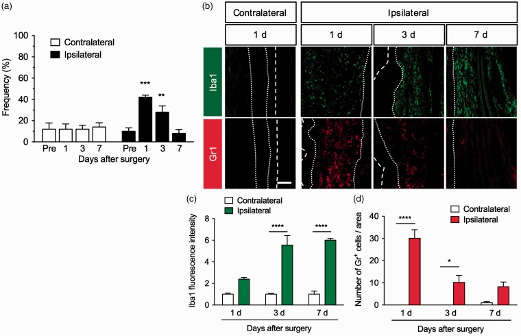
Figure 1.
Mechanical hypersensitivity and infiltration of macrophages and neutrophils around the incision site in a mouse model of postoperative pain. Plantar incision surgery was performed on the mouse right hind paw. (a) To assess mechanical sensitivities in the contralateral and ipsilateral hind paws, the frequency of paw withdrawal responses to von Frey filament stimulation was measured before the surgery (pre) as well as one, three, and seven days after the surgery. Data are expressed as means of the percentage ± S.E.M. n = 5. **P < 0.01, ***P < 0.001, compared with pre. (b) Representative photographs for immunofluorescence staining of Iba1 (green) and Gr1 (red) in the contralateral (one day) and ipsilateral hind paws one, three, and seven days after the surgery. Scale bar = 100 µm. White dashed lines mark the outer of epidermis. White dotted lines separate the dermis from the epidermis and fascia. Graphs show Iba1 fluorescence intensity (c) and the number of Gr1+ cells (d) in the contralateral and ipsilateral hind paws, n = 3. Data are expressed as means ± S.E.M. ****P < 0.0001, *P < 0.05.

