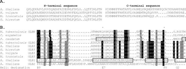
Figure 1.
The N- and C-terminal domains of some plant GLB3 proteins are shown in A, with a portion of the central region connecting them aligned with other Hbs in B. In A, the full C-terminal portion of cotton GLB3 is not shown, as it has not been determined. Shown in B is a structure-based sequence alignment of plant GLB3 proteins from A. thaliana, barley, barrel medic, and cotton, with 2-on-2 Hbs from microorganisms (2) and plant Hbs that have a myoglobin-like fold (11). Boxed regions denote α-helical regions in the structures of rice GLB1;1 and the 2-on-2 Hb of P. caudatum. Residues conserved between M. tuberculosis GLBO and GLB3 proteins are highlighted. The proximal (F8) and distal (E7) residues are marked with a star, and the helix designations for the beginning and end of the central domain are given.

