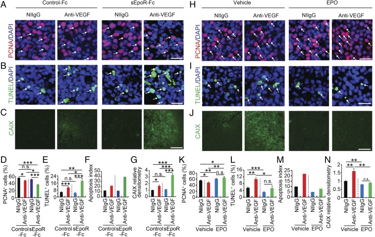
Fig. 4.
EPO inhibition increases antitumor activity by anti-VEGF drugs. (A–C) Immunohistochemical analyses of PCNA+ proliferating tumor cells, TUNEL+ apoptotic tumor cells, and tumor hypoxia. Arrows in A indicate PCNA+ proliferating tumor cells and arrows in B point to TUNEL+ apoptotic tumor cells. CAIX positive hypoxic signals are indicated with green signals in C. DAPI in blue was used for counterstaining of cell nuclei. (D–G) Quantification of PCNA+ proliferating tumor cells, TUNEL+ apoptotic tumor cells, apoptotic index, and CAIX positive hypoxic signals (n = 10 random fields per group; six animals per group). (H–J) Immunohistochemical analyses of PCNA+ proliferating tumor cells, TUNEL+ apoptotic tumor cells, and tumor hypoxia. Arrows in H indicate PCNA+ proliferating tumor cells and arrows in I point to TUNEL+ apoptotic tumor cells. CAIX-positive hypoxic signals are indicated with green signals in J. DAPI in blue was used for counterstaining of cell nuclei. (K–N) Quantification of PCNA+ proliferating tumor cells, TUNEL+ apoptotic tumor cells, apoptosis index, and CAIX-positive hypoxic signals (n = 10 random fields per group; six animals per group). *P < 0.05; **P < 0.01; ***P < 0.001; n.s., not significant. Data are means ± SEM. (Scale bars: A, B, H, and I, 50 μm; C and J, 100 μm.)

