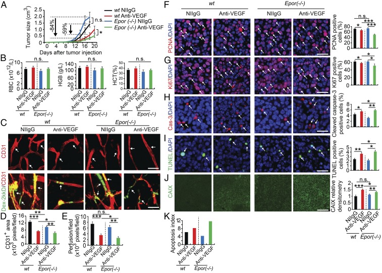
Fig. 5.
Nonhematopoietic ablation of EpoR increases anti-VEGF sensitivity. (A) Tumor growth rates (n = 6 animals per group). (B) Analyses of red blood cells, hemoglobin levels, and hematocrits (n = 6 animals per group). (C–E) Immunohistochemical analyses of CD31+ microvessels and vascular perfusion of 2,000-kDa dextran (n = 6 animals per group). Arrows in C, Upper point to CD31+ blood vessels and in Lower indicate perfused dextran+ signals (n = 10 samples per group). Quantifications of CD31+ vessel density and dextran blood perfusion (n = 10 samples per group). (F–K) Immunohistochemical analyses of PCNA+ proliferating tumor cells, Ki67+ proliferating tumor cells, cleaved caspase-3+ apoptotic tumor cells, TUNEL+ apoptotic tumor cells, and tumor hypoxia (n = 10 random fields per group; six animals per group). Epor (−/−) indicates EpoR(−/−)::HG1-EpoR mice. *P < 0.05; **P < 0.01; ***P < 0.001; n.s., not significant. Data are means ± SEM. (Scale bars: 50 μm.)

