Fig. 2.
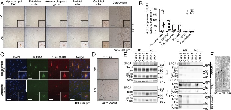
BRCA1 is mislocalized at the cytoplasm of AD brains, and it occurs at the insoluble fraction. (A) Immunohistochemical images of various regions from advanced-stage AD or NC by anti-BRCA1 antibody. Representative immunohistochemical images are shown from a total of n = 6 (hippocampus, entorhinal cortex, anterior cingulate gyrus, and parietal lobe) or n = 4 (occipital lobe and cerebellum) each. (B) Statistical analysis of A. Error bars represent means ± SEM. Significance was determined using one-way ANOVA followed by post hoc Holm–Sidak method. Mean ± SEM: NC: 3.72 ± 1.37/mm2 in the hippocampus, 1.35 ± 0.52/mm2 in the entorhinal cortex, 0/mm2 in the anterior cingulate gyrus, the parietal lobe, the occipital lobe, and the cerebellum. AD: 67.32 ± 21.27/mm2 in the hippocampus, 39.69 ± 8.05/mm2 in the entorhinal cortex, 15.33 ± 1.70/mm2 in the anterior cingulate gyrus, 8.46 ± 2.31/mm2 in the parietal lobe, 0.68 ± 0.48/mm2 in the occipital lobe, and 0/mm2 in the cerebellum. (C) Immunofluorescence images of advanced stage AD brains colabeled with DAPI, anti-BRCA1, and anti-pTau antibodies. Representative images from n = 4. (D) Immunohistochemical images of inferior temporal gyrus samples from NC or advanced-stage AD brains by anti–γ-H2ax antibody. Representative images are shown from a total of n = 3 samples of AD and NC. (E) Serial solubilization of postmortem brain samples using various detergents. The samples were treated with indicated detergents in a serial manner, each time saving the centrifuged supernatants as soluble fractions for each detergent. Three AD and NC samples were tested. (F) Immuno-EM of purified PHF by anti-BRCA1 antibody. *P < 0.05.

