Fig. 3.
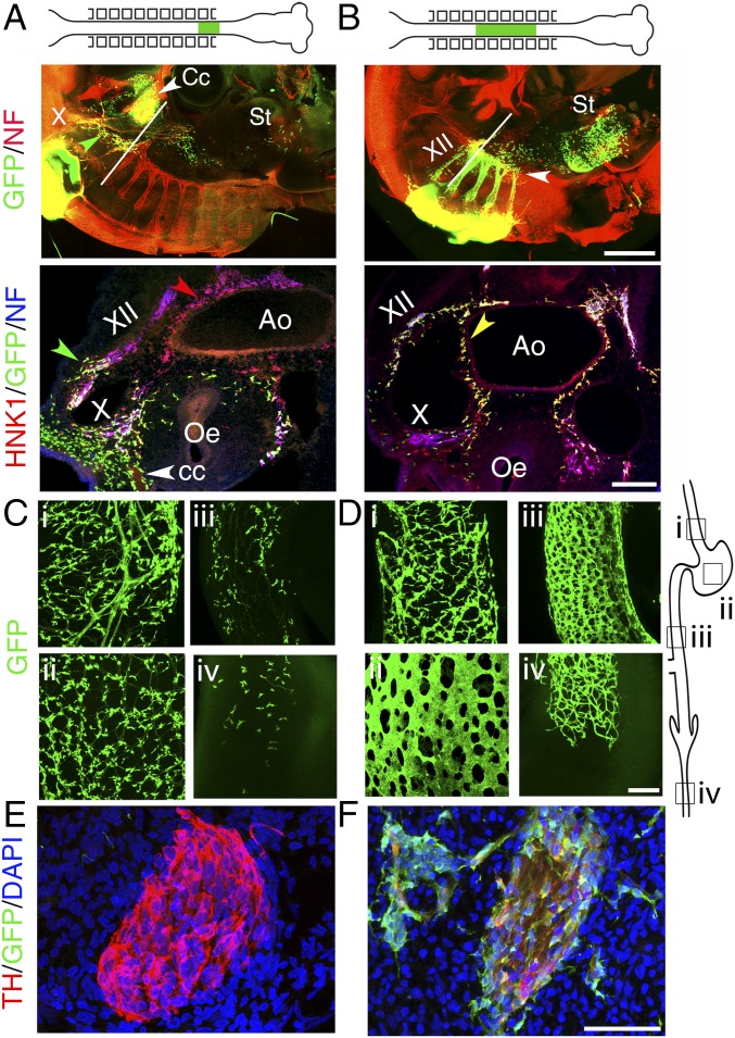
Two distinct streams of cells migrate into the esophagus in chicken embryos. (A) E3.5 chicken embryo after a stage 10 isotopic graft of the neural tube facing somites 1 and 2, from a GFP transgenic donor to a wild-type host. (Upper) Whole-mount lateral view showing that the graft has produced circumpharyngeal crest (Cc) and cells associated with fibers, mostly in the vagus nerve (X) but also in a connecting meshwork between the hypoglossal nerve and the nodose ganglion (green arrowhead); a few cells have migrated ahead of the vagus nerve, all of the way to the stomach (St). (Lower) Transverse section at the level indicated on the Upper panel, showing that the crest produced by the graft reaches the esophagus (Oe) by following the vagus, not the sides of the aorta (Ao) which is populated only by HNK1+;GFP− cells (red arrowhead). (B, Upper) Same as above but with a graft facing somites 3–7. The graft has produced cells associated with spinal nerves—and the hypoglossal (XII)—the nascent sympathetic chain (white arrowhead), a few cells in the esophagus and many more cells in the stomach than for the somite1-2 grafts. (Lower) Transverse section at the level indicated on the Upper panel, showing that the crest from the graft, apart from colonizing the hypoglossal (XII), follows the ventral path and reaches the esophagus by circumnavigating the dorsal aorta (yellow arrowhead), not by following the vagus. (C and D) Whole-mount views of the digestive tube at E7 showing the presence of graft-derived cells, after somite1–2 grafts (C) versus somite 3–7 grafts (D), at the rostro-caudal levels indicated in the schematic on the right: (i) esophagus; (ii) gizzard; (iii) preumbilical intestine; (iv) colon. At this stage the colon is still incompletely colonized (level iv). (E and F) Sagittal sections through the superior cervical ganglion at E5.5, stained with the indicated markers. [Scale bars: A and B (Upper), 500 µm; A and B (Lower), 100 µm; C–F, 50 µm.]

