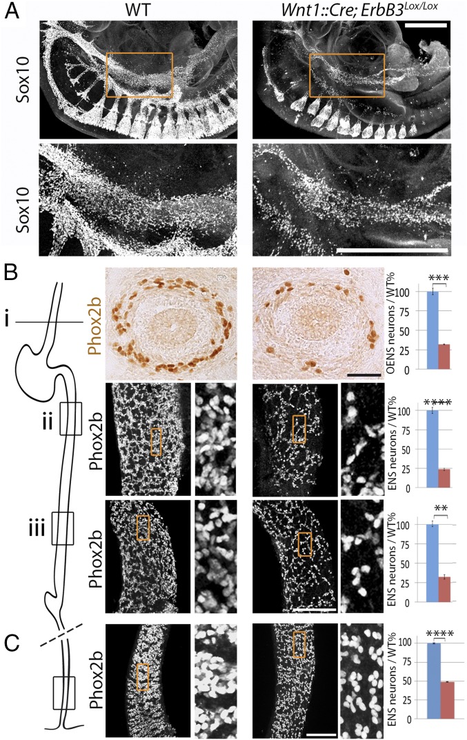
Fig. 4.
The ENS is atrophic in the absence of the tyrosine kinase receptor ErbB3. (A) Lateral views of wholemount E10.5 embryos stained for Sox10, in the indicated genotypes. In mutants where ErbB3 is deleted from the neural crest, Sox10+ cells are partially depleted in the foregut. (Lower) Magnified views of the area boxed in the Upper panels. (B) Sections through the esophagus (Upper) and whole mounts of the midgut at two rostro-caudal levels [(Lower) low magnifications at Left, enlarged views (7× zoom) of the boxed area at Right] indicated on the schematic on the Left, in E13.5 wild-type (Left column) and conditional ErbB3 mutants (Right column), stained for Phox2b. The atrophy of the enteric ganglia is quantified on the graphs. Wnt1::Cre;ErbB3lox/lox mutants showed fewer esophageal precursors (level i: 32 ± 0.6%/wild-type; P = 0.003, n = 3) and postgastric precursors (level ii: 24 ± 1.3%/wild-type; P = 0.0001, n = 4; level iii: 32 ± 2.8%/wild type; P = 0.007, n = 4). (C). Whole mounts of the distal hindgut (enlarged views of the boxed area on the Right), in E17.5 wild-type (Left column) and conditional ErbB3 mutants (Right column) stained for Phox2b (49 ± 0.41%; P = 0,00001, n = 5. (Scale bars: A, 500 µm; B and C, 100 µm.). Error bars indicate SEM. **P < 0.01, ***P < 0.005, ****P < 0.0005.

