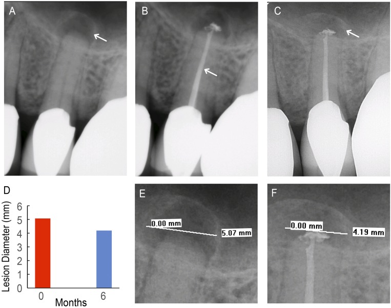
Fig. 4.
Control patient (C1). (A) Pretreatment radiograph of tooth 13 with existing apical lesion (white arrow). (B) Completed backfill (white arrow) and apical seal with unmodified GP. (C) Six-month follow-up radiograph shows the apical lesion with increased bone density (white arrow). (D) Lesion diameter at pretreatment (red) and 6-mo follow-up (blue) appointments. (E) A 5.07-mm apical lesion diameter was visible on the pretreatment radiograph. (F) A 4.19-mm apical lesion diameter was visible on the 6-mo follow-up radiograph.

