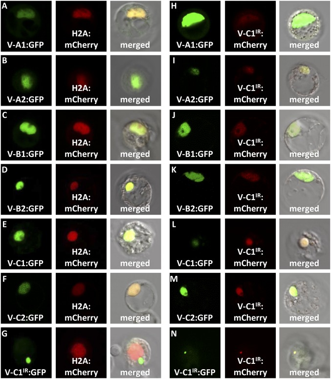
Fig. 2.
Subcellular colocalization demonstrates that PtrVND6-C1IR can be translocated from cytoplasmic foci into the nucleus. (A) PtrVND6-A1:GFP, (B) PtrVND6-A2:GFP, (C) PtrVND6-B1:GFP, (D) PtrVND6-B2:GFP, (E) PtrVND6-C1:GFP, and (F) PtrVND6-C2:GFP localized with H2A:mCherry in the nucleus, but (G) PtrVND6-C1IR:GFP localized in cytoplasmic foci. PtrVND6-C1IR:mCherry can be translocated into the nucleus by (H) PtrVND6-A1:GFP, (I) PtrVND6-A2:GFP, (J) PtrVND6-B1:GFP, (K) PtrVND6-B2:GFP, (L) PtrVND6-C1:GFP, and (M) PtrVND6-C2:GFP. (N) PtrVND6-C1IR:GFP and PtrVND6-C1IR:mCherry colocalized in cytoplasmic foci. The diameter of the SDX protoplasts is ∼30 μm.

