Fig. 2.
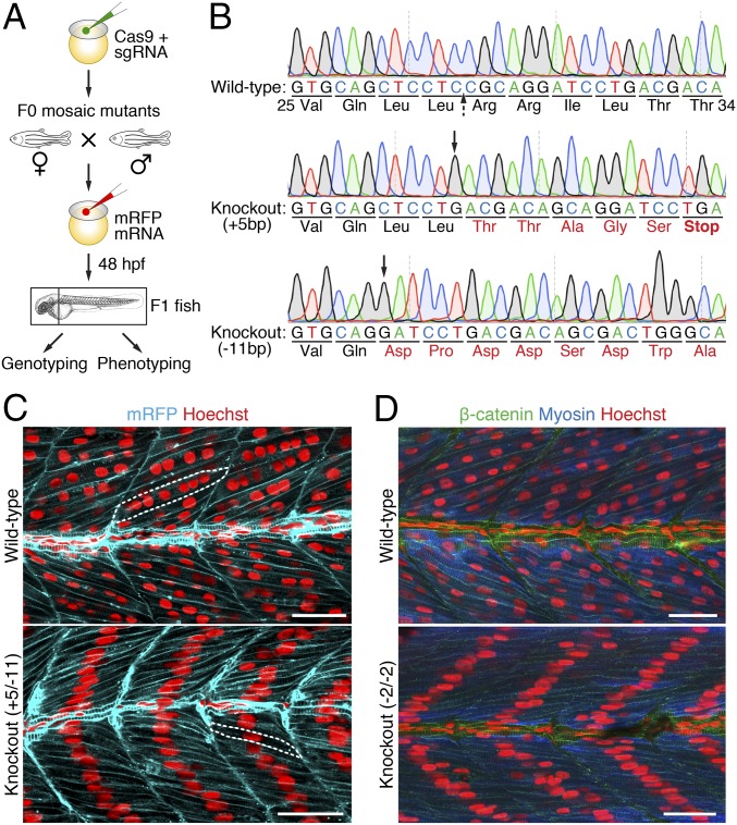
Ablation of myomixer in zebrafish abolishes myoblast fusion. (A) Schematic diagram of the experimental design. (B) DNA sequences of the myomixer coding region in wild-type and F1 knockout (KO) zebrafish. A region of wild-type (Val25–Thr34) and frame-shifted myomixer ORFs is shown. The Cas9 cleavage site is indicated by a dashed arrow, the starting positions of indels are indicated by black arrows. Amino acid mutations are in red. (C) Confocal images of 48-hpf wild-type and KO (+5/−11) embryos expressing a membrane-localized mRFP and stained with Hoechst to visualize the nuclei. Note the multinucleated muscle fibers (one of which is outlined) in the wild-type and the mononucleated muscle fibers (one of which is outlined) in the KO embryos. (D) Confocal images of 48-hpf wild-type and KO (−2/−2) embryos stained with anti–β-catenin, anti-fast muscle myosin, and Hoechst. (Scale bars, 25 µm.)

