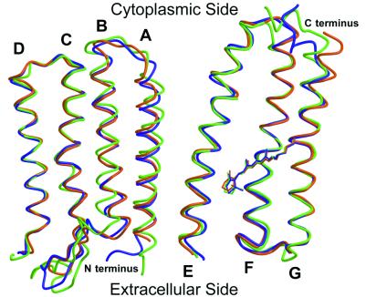
Figure 1.
Superposition of the Cα traces and the retinals of pSRII (orange), BR (9) (purple), and HR (14) (green). Helices C–G, which delineate the retinal-binding pocket, are structurally conserved among the three archaerhodopsins. Marked differences are visible in the BC loop and in the N and C termini. Figures were drawn with a modified version of molscript (27) and were rendered with RASTER 3D (28).

