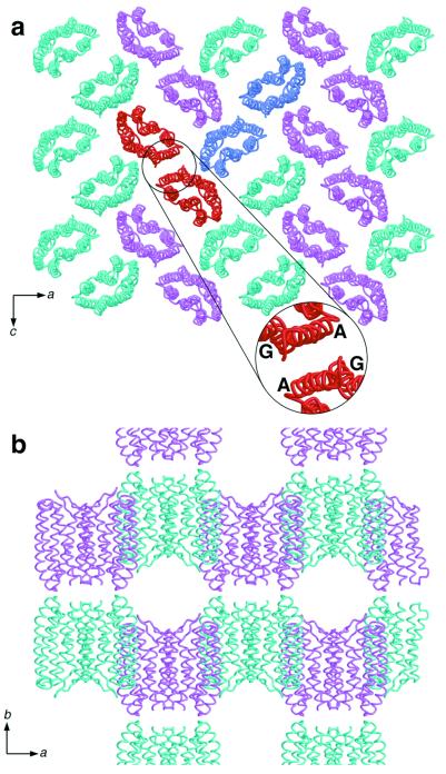
Figure 2.
Packing arrangement of pSRII. (a) Arrangement parallel to the ac plane, viewed along the b axis. Monomers are shown as cyan and pink ribbon models. The protein forms crystallographic dimers, and two dimers are highlighted in red and blue. (Inset) Dimer interface, consisting of helices A and G from both monomers. (b) Arrangement parallel to the ab plane viewed along the c axis showing the layer-like packing of pSRII molecules in the crystal.

