Figure 3.
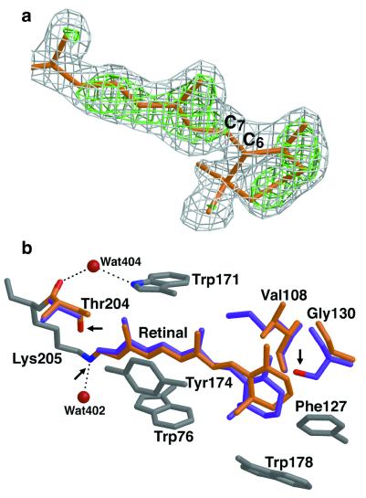
The retinal in pSRII. (a) Configuration of the β-ionone ring of the retinal. The 2Fobs−Fcalc (gray) and Fobs−Fcalc (green) electron-density maps contoured at 1.0 and 3.0 σ, respectively, were calculated before the inclusion of the retinal atoms into the model. They are superimposed on the refined model (orange). This view shows unambiguously that the retinal in pSRII is in the 6-s-trans configuration. (b) Comparison of the retinal-binding pockets in pSRII and BR. The retinal in pSRII (orange) is less bent than that in BR (purple). Conserved residues among archaerhodopsins are shown in gray. The nonconserved pSRII residues (Val-108, Gly-130, and Thr-204) are shown in orange, and the respective BR residues (Met-118, Ser-141, and Ala-215) are depicted in purple. Arrows indicate the Schiff-base nitrogen and the oxygens of Thr-204 in pSRII and of Ser-141 in BR. Modifications of residues at both ends of the retinal induce change of polarity in its binding pocket. Figures were drawn with a modified version of molscript (27) and were rendered with RASTER 3D (28).

