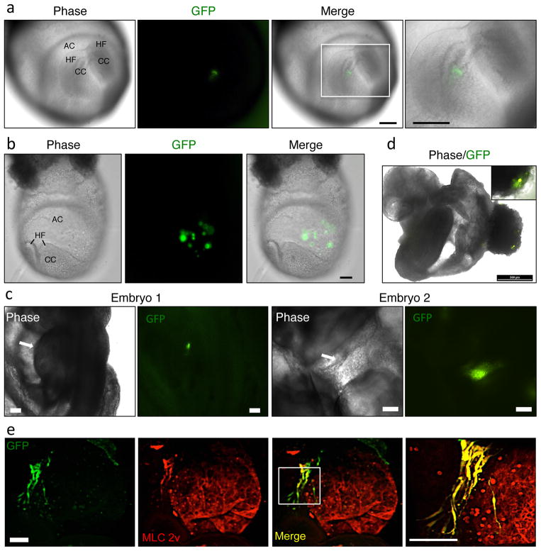
Figure 6. Injection of iCPCs into cardiac crescent for embryonic potency test.
(a) iCPCs labelled with GFP expressing lentivirus were injected into the cardiac crescent of mouse embryos. Images show a perfect injection where the cells are localized to the cardiac crescent. (b) Images show a faulty injection where the pipette pierced through the cardiac crescent and cells were injected into the amniotic cavity. (c) iCPCs injected into the cardiac crescent localize to the developing heart tube after 24 hr of whole embryo culture. Arrow indicates developing heart tube. (d) Adult cardiac fibroblasts injected into the cardiac crescent are excluded from the developing heart tube and localize to the ecto-placental cone (extra-embryonic tissue) after 24 hr of whole embryo culture. (e) iCPC-injected embryos were immunostained in whole-mount preparations for CM markers and GFP. Three-dimensional reconstruction images show iCPCs differentiated into CMs, as indicated by co-expression of CM marker MLC-2v and GFP. AC=amniotic cavity, HF=head fold, CC=cardiac crescent. Scale bar 100um in a,b,c,e. 500 um in d. All animal experiments were performed in accordance to University of Wisconsin-Madison’s animal use guidelines.

