
Fig. 1.
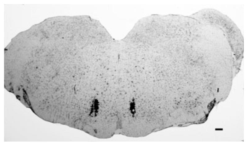
A representative photomicrograph of a medullary section at the level of the rostroventromedial medulla (RVM) counterstained with Nissl stain. The microinjection sites in the RVM were visualized with the microinjection of India ink to verify proper cannula placement. Scale bar: 100 μm.