
Fig. 2. Anchored PKA holoenzyme dynamics upon ligand stimulation.
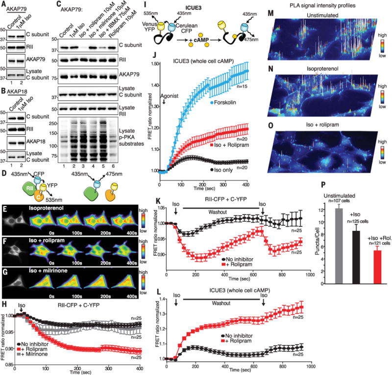
Western blot analysis of PKA C and RII subunits in (A) AKAP79 or (B) AKAP18g immunoprecipitates (IPs) after isoproterenol stimulation (Iso). (C) IP and Western blot analysis of AKAP79 complexes isolated in the presence of phosphodiesterase inhibitors. (D) Schematic depicting RII-CFP and C-YFP intermolecular FRET. (E to H) FRET analysis of holoenzyme dissociation in HEK293 cells. (E) Time course of representative cells (0 to 400 s) stimulated with isoproterenol. (F) Iso + PDE4 inhibitor rolipram. (G) Iso + PDE3 inhibitor milrinone. (H) Amalgamated data from 25 recordings under each experimental condition. (I) Schematic depicting ICUE3 cAMP FRET sensor. (J) ICUE3 FRET recordings show cAMP production in response to Iso (black), Iso + rolipram (red), and forskolin (blue). The number of experiments is indicated. (K and L) FRET analysis upon two trains of hormonal stimulation. (K) Intermolecular FRET analysis of holoenzyme dissociation in HEK293 cells upon two trains of stimulation with isoproterenol (black) and Iso + PDE4 inhibitor rolipram (red). (L) Monitoring cAMP accumulation with ICUE3 FRET sensor in response to isoproterenol (black) and Iso + PDE4 inhibitor rolipram (red). Amalgamated data from 25 recordings under each experimental condition. (M to O) Proximity ligation assay (PLA) signal-intensity projections show RII-C interactions in (M) Unstimulated, (N) Iso-stimulated, and (O) Iso- and rolipram-treated HEK293 cells. (P) Quantification of PLA puncta per cell using Fiji/ImageJ. All data are presented as means ± SEM.